



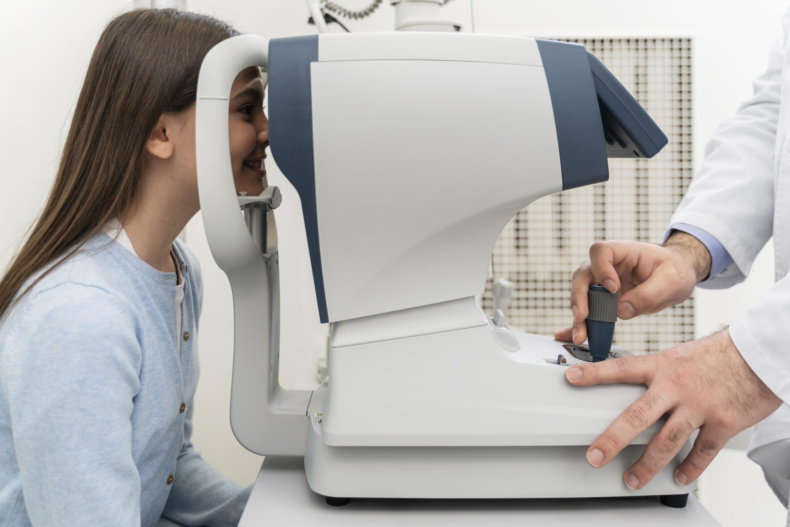
Eye care specialists diagnose RVO through an eye exam and retinal imaging tests. They also coordinate care with your primary care physician (PCP) to discover the cause of blood flow problems.
Eye exam
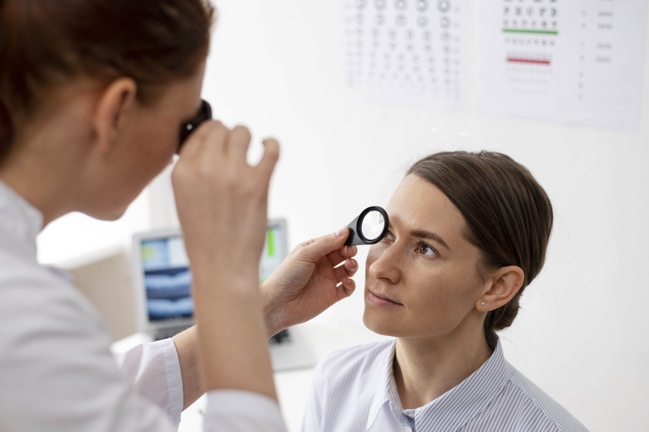
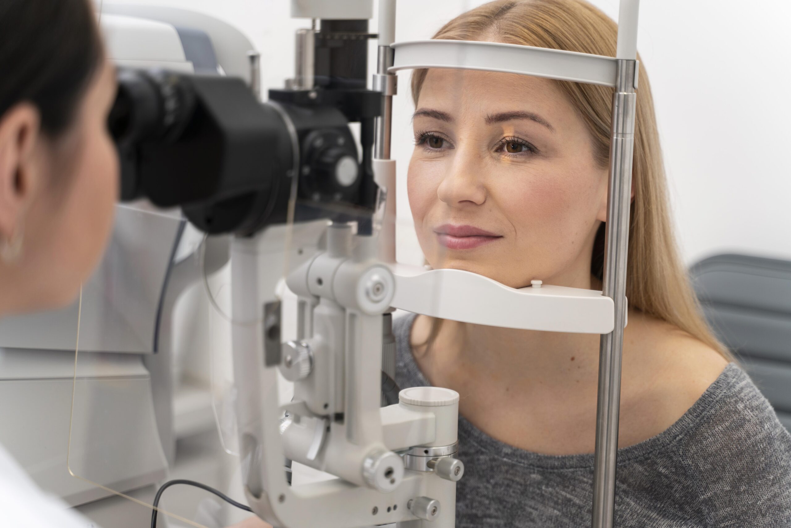
Your eye care specialist will dilate your pupils so they can see into the back of each eye. They’ll use a microscope and a head-mounted ophthalmoscope to shine a light into your eye. They’ll closely examine the inside of your eye to look for complications and signs of vision loss.
This exam can help:
- Distinguish between central and branch RVO.
- Identify signs of macular edema and abnormal blood vessel formation.
- Estimate how much of your retina lacks blood flow.
You may need further testing to diagnose RVO and show the extent of complications.
Tests to diagnose retinal vein occlusion
Your eye care specialist may use one or more of the following tests to help diagnose and describe your condition:
- Fundus photography: This form of retinal imaging shows the presence of abnormal new blood vessels and the amount of bleeding inside your eye.
- Optical coherence tomography (OCT): This form of high-resolution imaging shows the presence of macular edema. It measures the thickness of your retina and provides precise numbers that help guide the treatment of your condition over time.
- Fluorescein angiography: For this test, your provider injects dye into a vein in your arm. The dye travels to the blood vessels in your retina and makes them stand out in imaging. Your provider may use this form of imaging to show the extent of a blockage in your retinal vein. This test also shows how much of your retina isn’t receiving adequate blood flow.
Coordinated care Your eye care specialist and primary care physician will work together to find the cause of RVO and lower your risk for future issues. You may need blood tests to check your cholesterol levels, blood sugar and other important numbers.