


Keratoconus
If your cornea changes shape and becomes more like a cone than a dome, your vision is likely to be less sharp. Treatments range from glasses or contact lenses, cross linking to a cornea transplant. In most cases, keratoconus has no definite cause


Overview
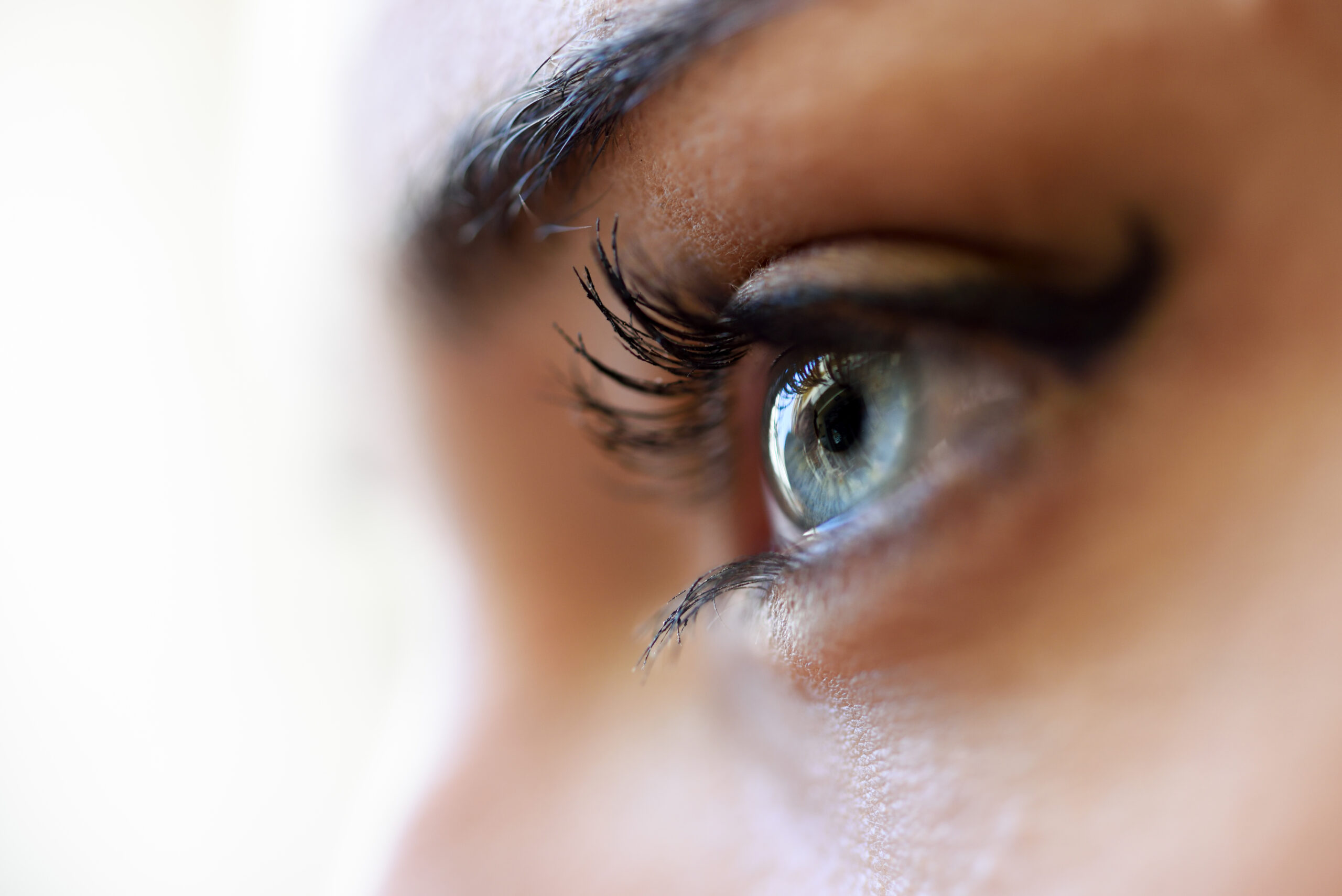
The rounded cornea protects the lens and iris in your eye, but if you have keratoconus, your cornea is more pointed like a cone.
What is keratoconus?
Keratoconus is a condition of the eye in which the normally rounded cornea bulges outward into a cone shape. The cornea is the clear, central part of the front surface of the eye. It protects your eye and helps you focus for clear vision. You pronounce keratoconus as care-ah-ta-KO-nus.
Eye care providers normally find keratoconus during your teenage years or your 20s and 30s, but it can also start in childhood. In some cases, a provider will diagnose a mild case of keratoconus at a later age. The changes in the shape of the cornea occur over several years but happen at a more rapid rate in younger people.
How does keratoconus affect your vision?
Keratoconus changes vision in two ways:
- As the cornea changes to a cone shape, the smooth surface also warps. The term for this is irregular astigmatism. Glasses can’t fully correct irregular astigmatism.
- As the front of the cornea steepens, your eye becomes more nearsighted. As a result, you may need new glasses more often.
How common is keratoconus?
One estimate says that keratoconus develops in 50 to 200 of every 100,000 people.
Symptoms and Causes
What are the Keratoconus symptoms?
The main symptoms of keratoconus include:
- Gradually worsening vision in one or both eyes. (Generally, keratoconus affects both eyes.)
- Having double vision when you look out of just one eye.
- Seeing halos around bright lights.
- Being sensitive to light. (The term for this sensitivity is photophobia.)
- Having vision that gradually becomes distorted. With distorted vision, straight lines might look curvy or bent, and objects don’t have their correct shape.
What causes keratoconus?
The cause of keratoconus is largely unknown. Some studies have found that keratoconus runs in families, and that it happens more often in people who have certain medical conditions.
In most cases, you don’t have an eye injury or a disease that leads to keratoconus. People with keratoconus tend to rub their eyes a lot, which may cause the condition to develop more rapidly.
What conditions may be associated with keratoconus?
Keratoconus has a link to certain conditions that may also be associated with chronic eye rubbing. These conditions include:
- Atopic dermatitis and allergic dermatitis.
- Allergic rhinitis.
- Asthma.
- Down syndrome.
- Ehlers-Danos syndrome.
- Osteogenesis imperfecta.
- Congenital (present at birth) disorders that affect your eyes, like aniridia.
What are the complications of keratoconus?
Potential complications of keratoconus include:
- Corneal scarring: Scars develop on the cornea
- Corneal hydrops: Your cornea swells up with fluid suddenly.
- Fleischer rings: Iron deposits cause rings within your cornea.
- Vision loss: Vision loss can be mild or severe.
Diagnosis and Tests
What is keratoconus diagnosis?
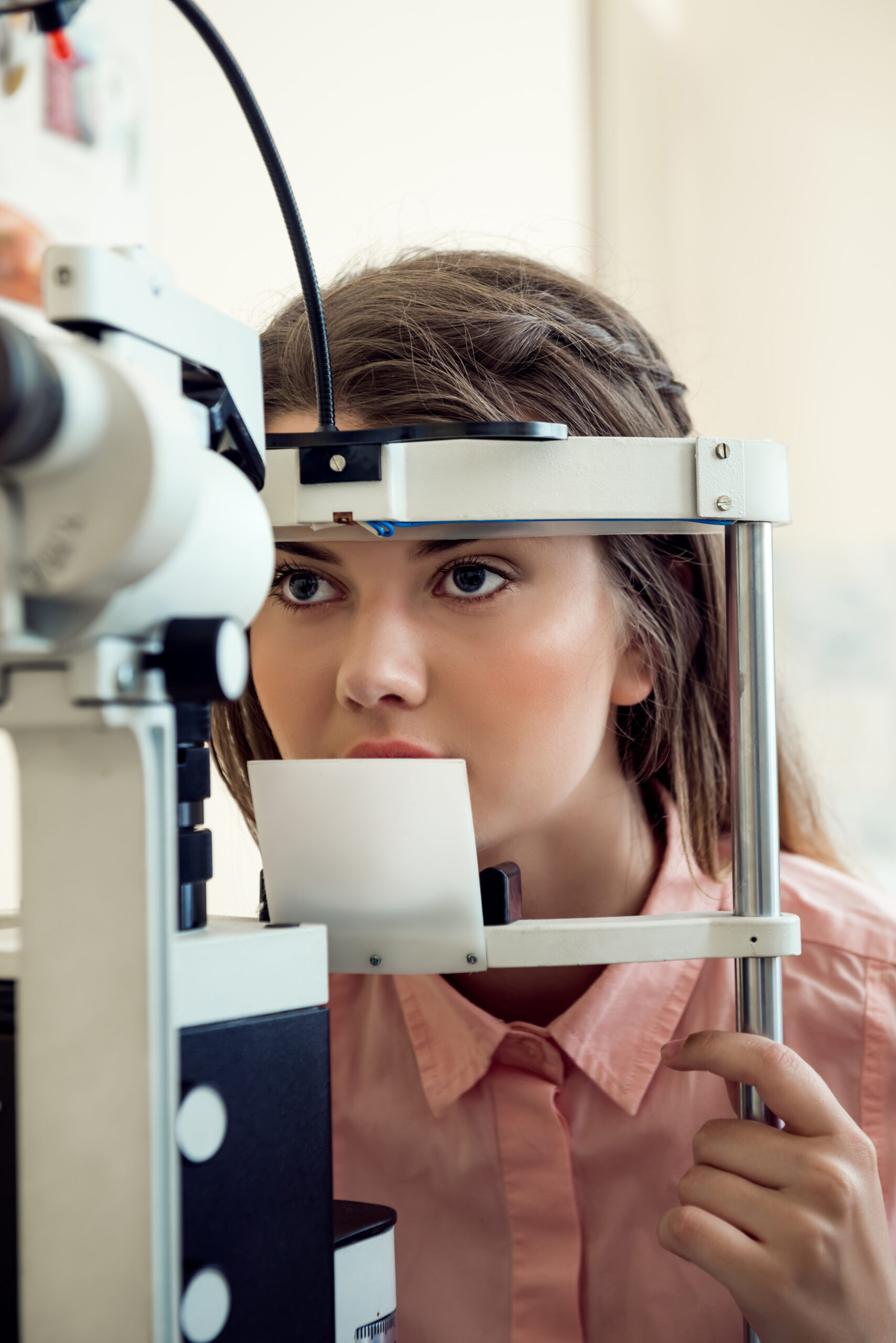
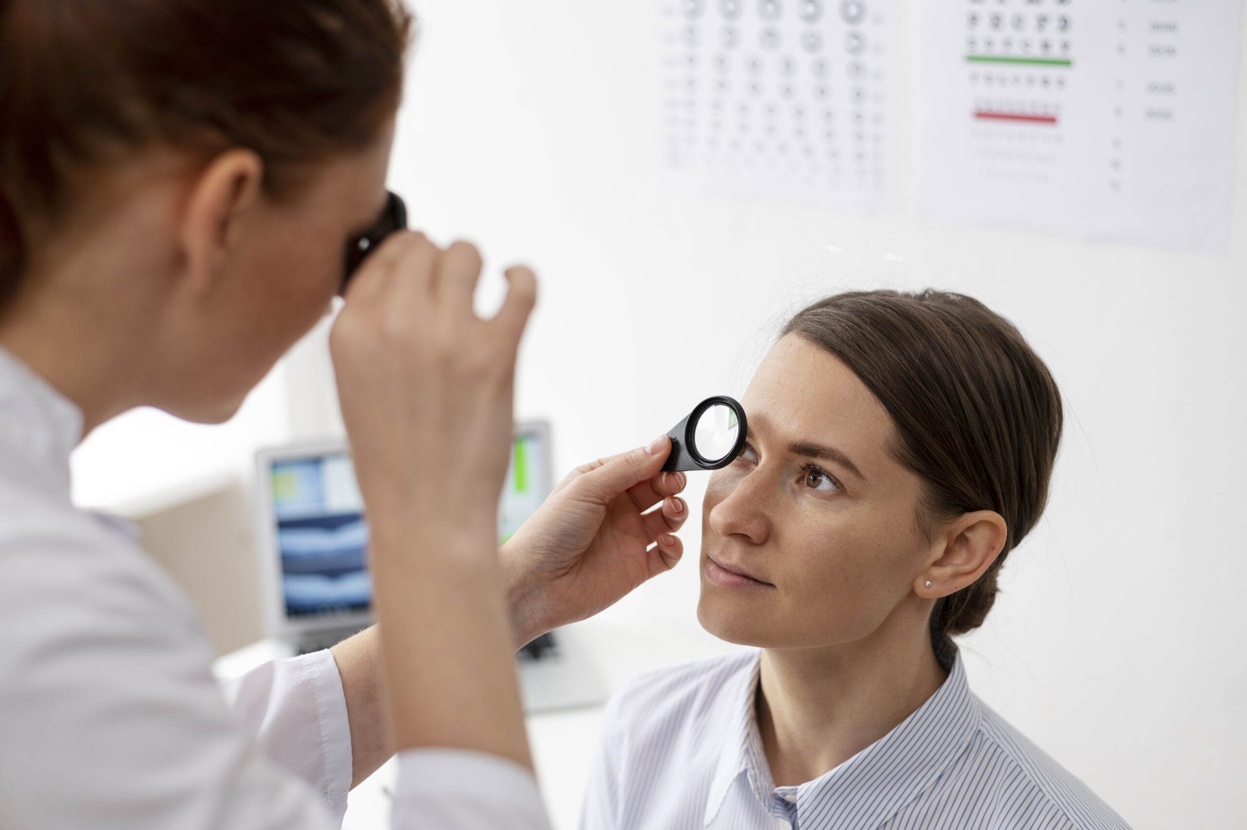
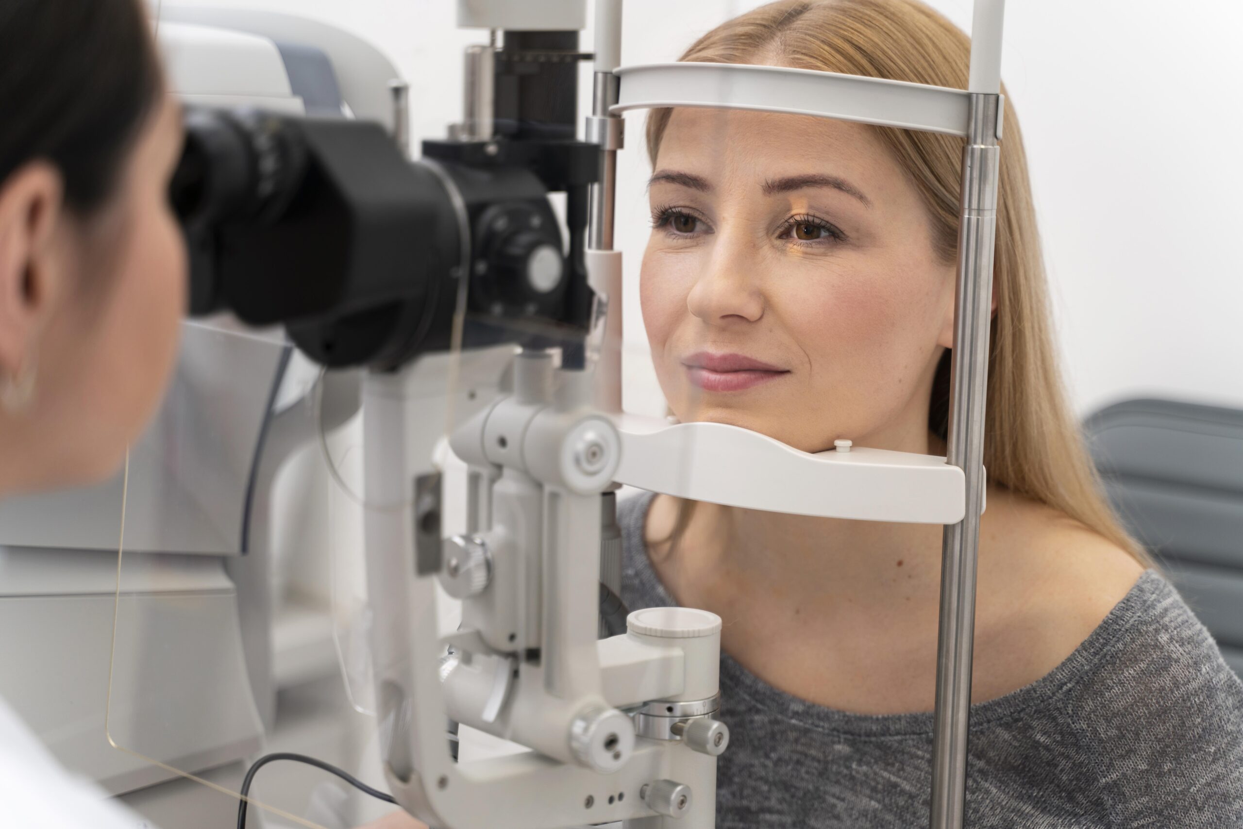
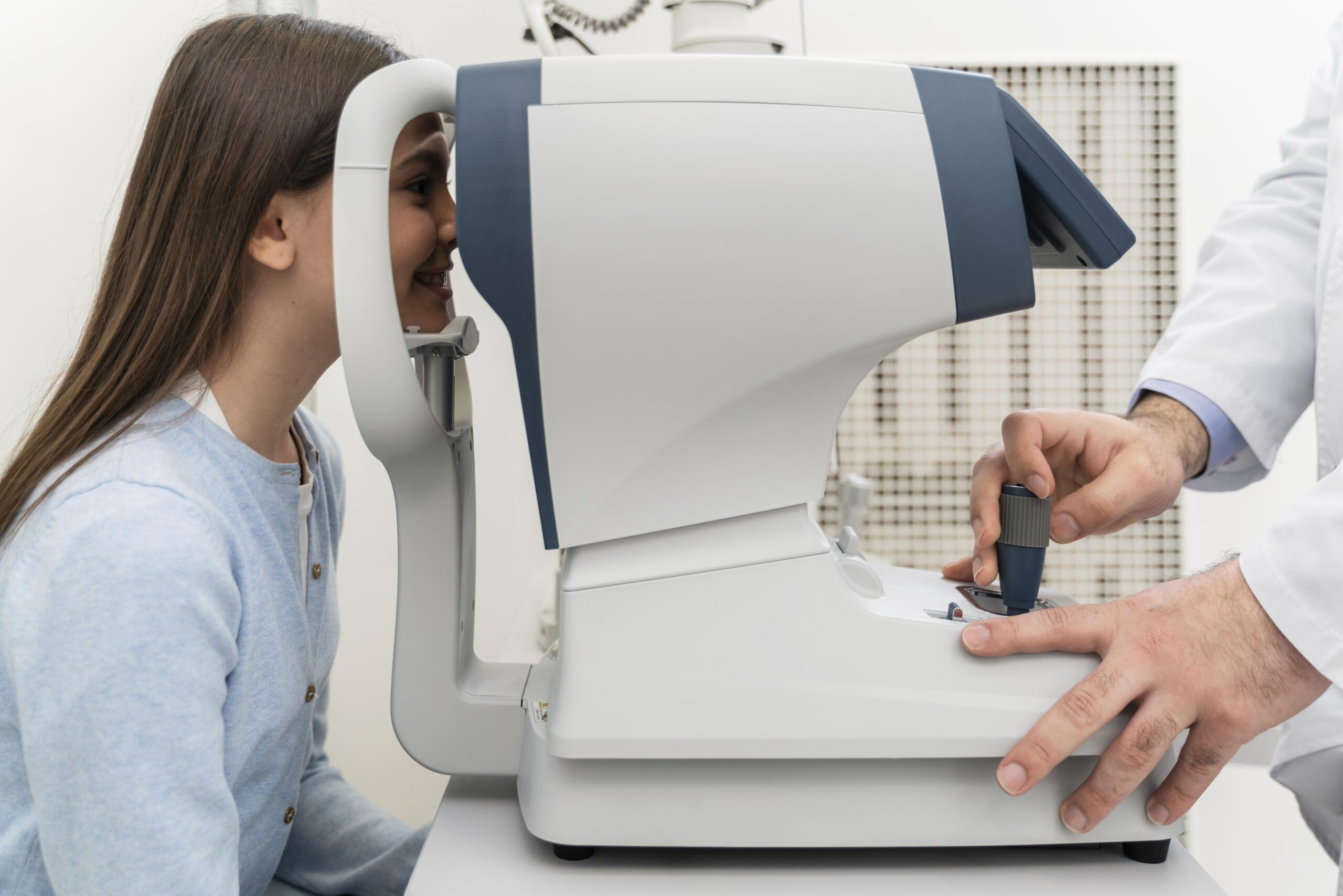
Your eye care provider will begin by asking you about your medical and family history and your symptoms. They’ll then do a comprehensive eye exam. They may also order one or more of the following tests:
- Visual acuity: This exam helps you find your correct prescription for clear vision.
- Slit lamp examination: This test combines a bright light and a microscope.
- Keratometry: This test measures corneal shape and astigmatism.
- Corneal mapping (tomography and topography): These tests measure the curve of the surface of your eye and produce a “map” of your cornea. Keratoconus shows up on these tests.
Management and Treatment
How is keratoconus treated?
There are several methods for treating keratoconus, depending on how severe the condition is. Your eye care provider can help to decide which, if any, of these treatments may help you. Treatments include eyeglasses, contact lenses, implantable ring segments, corneal crosslinking and cornea transplant.
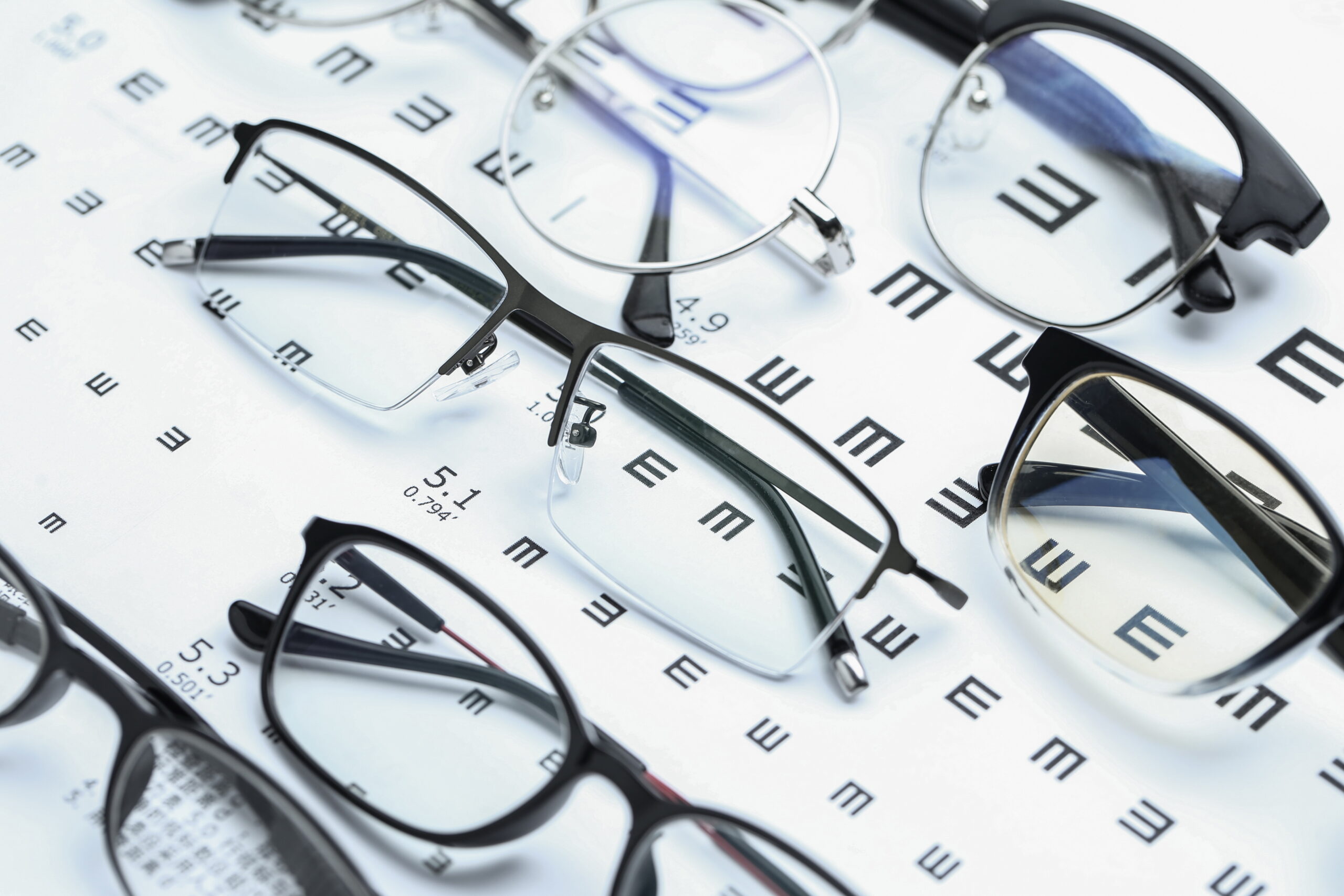
Eyeglasses and contact lenses for keratoconus
In the early stages of the disease, you can correct vision with normal eyeglasses or soft contact lenses. As keratoconus gets worse, eyeglasses may not correct your vision because of the amount of irregular astigmatism. You may need a special type of hard contact lens.
Corneal crosslinking to treat keratoconus
Corneal crosslinking uses ultraviolet (UV) light treatment that may slow or stop the keratoconus from getting worse. In this procedure, you also receive local anesthesia. Your provider puts drops of a drug containing riboflavin (vitamin B2) into your eye for up to 30 minutes. Then, your provider exposes your eye to an UV light for up to 30 minutes. The purpose of corneal crosslinking procedure is to strengthen the bonds between your cornea’s collagen fibers and surrounding proteins. This can help keep your cornea’s shape from getting steeper.
Implantable ring segments (INTACS) to treat keratoconus
INTACS® are small devices your provider inserts into your cornea to improve vision or make it easier to fit you with contact lenses. Your provider performs this procedure while you’re under local anesthesia (drops numb your eye). Then, your provider creates channels in the cornea and inserts the rings into these channels. The rings help to flatten the cornea and partially correct the cone shape the keratoconus causes.
Cornea transplant for keratoconus
Your provider may suggest that you have a cornea transplant if you have keratoconus that’s advanced. They’ll replace your diseased cornea with corneal tissue from a human donor. Usually, people with keratoconus have better vision after the transplant, but it may take more than a year for vision to stabilize. Some people may still need a specialty contact lens after the transplant to get their best vision.
Living With
When should I see my healthcare provider?
You should always contact your eye care provider when you have changes in vision. If you have keratoconus, you’ll probably need to see your provider on a regular basis. Keep your appointments.
When should I go to an emergency room?
Go to an emergency room (ER) if:
- You have a sudden loss of vision.
- You have severe eye pain.
- You’ve recently had an eye procedure and now have signs of infection, like a fever or discharge from your eye.